**What's it all about?**

We want you to tell us if you think the bacteria that you see is resistant or sensitive to the antibiotic ciprofloxacin. In this workflow you will see images of *Escherichia coli* (*E. coli*) that we took on a laboratory microscope.


**What does resistance look like?**

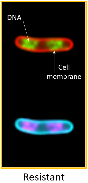
When a bacterium is resistant to the effects of ciprofloxacin, its DNA will  be more spread out (diffuse), throughout the bacterium, and there might be multiple copies of the DNA.
In all of the images you will see, the bacterial DNA will be shown in green and the cell membrane will be shown in red.

**Examples of a resistant *E.coli***
**What does sensitivity look like?**

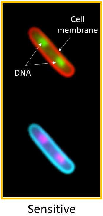
When a bacterium is sensitive to ciprofloxacin, the DNA will break when the cell tries to make more DNA in order to divide. In a sensitive bacterium, the DNA will collapse into a smaller, denser ball.

**Examples of a sensitive *E.coli***
**Be careful...**
All of these images are just a snapshot of the cell's response. The images are captured after 30 minutes of antibiotic treatment.

In some cases, there may be sensitive cells where the DNA is not yet completely condensed. In the example below, you can see a sensitive cell that still has two distinct clumps of DNA, but, the DNA in each clump is more compact than in the resistant example.
*If you are unsure whether a bacterium looks like it is resistant or sensitive to ciprofloxacin, try to give a best guess based on how compact or not the DNA looks.*
System and task instructions
System instruction
Task instruction
Is this bacterium resistant or sensitive to the antibiotic?